Unlock hundreds more features
Save your Quiz to the Dashboard
View and Export Results
Use AI to Create Quizzes and Analyse Results

Sign inSign in with Facebook
Sign inSign in with Google

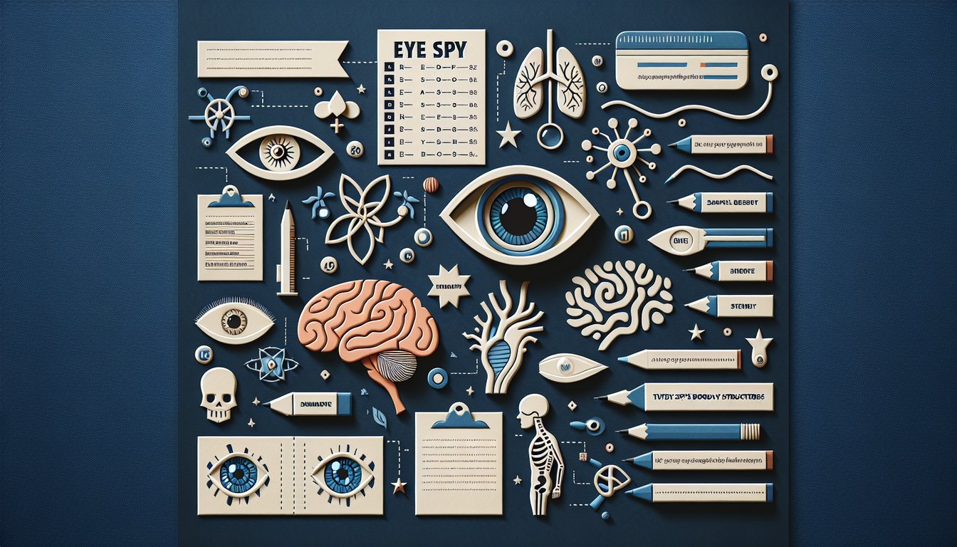
Practice Quiz: Identify Eye Structures

Test Your Skills on Eye Anatomy Today

Difficulty: Moderate
Grade: Grade 8
Study OutcomesCheat Sheet
Colorful paper art promoting Eye Spy Anatomy quiz for high school students.

What is the colored part of the eye?
Retina
Cornea
Sclera
Iris
The iris is the colored part of the eye and controls the amount of light entering by adjusting the pupil size. Its ability to change shape is key to regulating light exposure on the retina.
Which structure is the clear, dome-shaped surface covering the front of the eye?
Lens
Iris
Pupil
Cornea
The cornea is the transparent, dome-shaped outer layer that covers the front of the eye. It helps focus incoming light onto the lens and retina.
What is the small opening in the center of the iris called?
Pupil
Lens
Cornea
Sclera
The pupil is the dark, central opening in the iris that permits light to enter the eye. It adjusts size based on light conditions, controlling light exposure.
Which structure is a transparent, flexible component that focuses light on the retina?
Cornea
Lens
Iris
Vitreous Humor
The lens is responsible for bending (refracting) light to focus it precisely on the retina. Changes in its shape allow the eye to accommodate objects at different distances.
Which part of the eye is responsible for detecting light and converting it into neural signals?
Retina
Sclera
Iris
Cornea
The retina is a layer of light-sensing cells at the back of the eye that converts light into electrical signals. These signals are then transmitted to the brain via the optic nerve.
What is the primary function of the sclera?
It refracts light onto the retina.
It produces aqueous humor.
It provides structural support and protects the inner eye.
It transmits visual signals to the brain.
The sclera is the white, fibrous outer layer of the eye that maintains its shape and protects delicate internal structures. Its toughness helps safeguard the eye.
What is the main function of the choroid?
It controls the size of the pupil.
It supplies oxygen and nutrients to the retina.
It focuses light onto the retina.
It produces vitreous humor.
The choroid is a layer rich in blood vessels that nourishes the outer layers of the retina. Its vascular nature is essential for the retina's health and function.
In which part of the eye is the aqueous humor located?
Anterior chamber
Scleral layer
Posterior chamber
Vitreous chamber
Aqueous humor is the clear fluid filling the space between the cornea and the iris, known as the anterior chamber. This fluid maintains intraocular pressure and provides nutrients to the eye.
What role does the vitreous humor serve in the eye?
It provides blood supply to the retina.
It maintains the shape of the eye and acts as a light-transmitting gel.
It refracts light to focus images on the retina.
It controls the size of the pupil.
The vitreous humor is a clear, gel-like substance that fills the space between the lens and the retina, helping maintain the eye's spherical shape. It allows light to pass through to the retina with minimal distortion.
How does the iris regulate the amount of light entering the eye?
By changing the shape of the lens.
By adjusting the size of the pupil.
By refracting light toward the retina.
By secreting aqueous humor.
The iris functions like a camera aperture by contracting or dilating to control the size of the pupil. This regulation adjusts the amount of light that reaches the retina.
What role does the fovea centralis play in vision?
It provides sharp, central vision with a high density of photoreceptors.
It controls the eye's peripheral vision.
It is responsible for color detection in low light.
It produces the vitreous humor.
The fovea centralis is a specialized area within the retina that is densely packed with cone cells. This concentration allows for detailed and high-resolution central vision.
What is the main function of the optic nerve?
It protects the retina from excess light.
It adjusts the shape of the lens for focusing.
It transmits visual information from the retina to the brain.
It regulates the production of aqueous humor.
The optic nerve is the conduit that carries electrical impulses from the retina to the brain, allowing visual information to be processed. Without it, the brain would not receive images from the eye.
Which structure produces the aqueous humor?
Iris
Cornea
Ciliary body
Lens
The aqueous humor is produced by the ciliary body, which is located behind the iris. This fluid is essential for maintaining intraocular pressure and nourishing the eye.
What role does the ciliary muscle play in focusing the eye?
It controls the size of the pupil.
It produces the vitreous humor.
It adjusts the shape of the lens for accommodation.
It refracts light as it enters the eye.
The ciliary muscle adjusts the curvature of the lens, a process known as accommodation, to ensure that images are in focus. This adjustment allows clear vision at both near and far distances.
What defines the anterior chamber of the eye?
The outer protective layer of the eye.
The fluid-filled space between the cornea and the iris.
The space between the lens and the retina.
The inner core of the eye where the vitreous humor is located.
The anterior chamber is the space at the front of the eye, filled with aqueous humor, located between the cornea and the iris. This chamber helps maintain intraocular pressure and provides nutrients to the eye's structures.
How does the structure of the cornea contribute to its role in focusing light?
Its curved, transparent surface refracts and directs light onto the retina.
Its flat surface minimizes light refraction for accurate imaging.
Its thick composition prevents any light from scattering.
Its opaque, vascular nature absorbs excess light.
The cornea's curved and clear structure is fundamental to its ability to refract incoming light effectively. This refractive power helps to focus light sharply onto the retina for clear vision.
What is the function of the blood-retinal barrier in eye anatomy?
It enhances blood flow to the retina by dilating retinal vessels.
It prevents harmful substances from entering the retina while allowing essential nutrients to pass through.
It aids in the drainage of aqueous humor to maintain intraocular pressure.
It facilitates rapid exchange of large molecules between the blood and retinal cells.
The blood-retinal barrier is vital for protecting the delicate neural tissues of the retina by regulating the passage of substances. It ensures that only necessary nutrients reach the retina while blocking potentially harmful agents.
Why is the fovea centralis particularly crucial for tasks requiring fine visual detail?
Because it increases the overall light sensitivity of the eye in low light conditions.
Because it contains more rod cells, which are essential for color vision.
Because it is responsible for peripheral vision and detecting motion.
Because it has a high concentration of cone cells, allowing for sharp, detailed central vision.
The fovea centralis, being densely packed with cone cells, is specialized for high-acuity central vision. This makes it essential for activities like reading or recognizing fine details.
How would damage to the optic nerve likely impact vision?
It might lead to an increase in intraocular pressure.
It could result in partial or complete vision loss in the affected eye.
It would primarily affect the eye's ability to focus on close objects.
It would only disturb the perception of colors without affecting overall vision.
The optic nerve is essential for transmitting visual information from the retina to the brain. Damage to it disrupts this pathway, leading to significant visual impairment or loss.
What is the relationship between the ciliary body and the process of accommodation, and why is it important?
The ciliary body modifies the curvature of the cornea to improve image focus.
The ciliary body controls the size of the pupil during accommodation.
The ciliary body, through the ciliary muscle, adjusts the shape of the lens to focus on objects at different distances.
The ciliary body produces aqueous humor, which indirectly influences lens transparency.
Accommodation is the process by which the eye changes its focus from distant to near objects, and this is achieved by altering the lens shape. The ciliary body, by contracting the ciliary muscle, facilitates this change in lens curvature, which is critical for clear vision.
0
{"name":"What is the colored part of the eye?", "url":"https://www.quiz-maker.com/QPREVIEW","txt":"What is the colored part of the eye?, Which structure is the clear, dome-shaped surface covering the front of the eye?, What is the small opening in the center of the iris called?","img":"https://www.quiz-maker.com/3012/images/ogquiz.png"}

Study Outcomes

  1. Identify key anatomical structures of the eye.
  2. Describe the function of each eye component.
  3. Analyze the relationships between different eye structures.
  4. Apply anatomical knowledge to evaluate eye-related questions.
  5. Synthesize information to reinforce understanding of eye anatomy.

Eye Anatomy Quiz: Identify Structures Cheat Sheet

  1. Primary structures of the eye - Your eyeball superstar team includes the cornea, lens, retina, iris, and optic nerve, each with a special gig from bending light to firing visual fireworks at your brain. Understanding these parts is like unlocking cheat codes for perfect vision. nursestudy.net
  2. Photoreceptor functions - Rods and cones are your retina's night‑vision and color squad: rods let you see in low light in shades of gray, while cones bring the rainbow party when the sun is out. Get to know these cells to ace questions on how you adapt from dusk to dawn. picmonic.com
  3. Extraocular muscles - Meet the six tiny engineering marvels: medial rectus, lateral rectus, superior rectus, inferior rectus, superior oblique, and inferior oblique. These muscles work in tandem like a choreographed dance crew to swivel your eyeballs in every direction. dailymeded.com
  4. "LR6SO4O3" mnemonic - This catchy code helps you remember that the lateral rectus is cranial nerve VI, superior oblique is IV, and the other four muscles are served by III. It's the quickest brain hack to avoid cranial nerve confusion during exams. pacs.de
  5. Miosis vs. Mydriasis - Miosis is your pupil's squeeze play (constriction) and mydriasis is the big stretch (dilation) - always remember mydriasis has a "D" for dilation. A quick mnemonic keeps you from mixing up these tiny tweaks. medicowesome.com
  6. Iris regulation - Think of the iris as your personal camera aperture, flexing its muscles to adjust pupil size and let in just the right amount of light. This dynamic control keeps your vision crisp whether you're in a dim dungeon or a sunny beach. opticianstudy.com
  7. Accommodation mechanism - When you switch focus from a distant mountain to your phone screen, the lens changes shape thanks to the ciliary muscles - like a zoom lens in your eye. Mastering this process will make questions on near and far vision a breeze. opticianstudy.com
  8. Optic nerve pathway - The optic nerve is the superhighway that carries image data from your retina to the visual cortex, where your brain builds the final picture. Recognizing this route is key to understanding vision disorders and visual field defects. opticianstudy.com
  9. Anatomical orientation - Medial means toward your nose, lateral is away, superior points up, and inferior points down - a simple compass for describing eye movements. Nail these terms to precisely document and interpret ocular exams. opticianstudy.com
  10. Eye layer structure - The eye's layers include the tough, white sclera on the outside, the vascular choroid in the middle, and the sensory retina lining the inside with photoreceptors. Knowing each layer's role is essential for spotting pathologies and layering your knowledge. opticianstudy.com
Powered by: Quiz Maker