Unlock hundreds more features
Save your Quiz to the Dashboard
View and Export Results
Use AI to Create Quizzes and Analyse Results

Sign inSign in with Facebook
Sign inSign in with Google

Master the Auditory System Anatomy Quiz

Discover Ear Anatomy with Interactive Questions

Difficulty: Moderate
Questions: 20
Learning OutcomesStudy Material
Colorful paper art depicting various elements of the auditory system for an anatomy quiz

Ready to explore ear anatomy and refine your understanding of the auditory pathway? This Auditory System Anatomy Quiz offers 15 multiple-choice questions designed for anatomy students, audiology trainees, and science enthusiasts. Test your knowledge of ear structure and sound transmission through engaging challenges, then customise questions in the editor to suit your study needs. You can even compare your performance with our Anatomy and Physiology Knowledge Quiz or revisit fundamentals in the Anatomy Fundamentals Quiz. Dive into more quizzes to keep mastering anatomy today.

Which part of the ear is primarily responsible for capturing sound waves and funneling them into the ear canal?
Cochlea
Pinna (auricle)
Tympanic membrane
Eustachian tube
The pinna, or auricle, is the visible outer part of the ear that captures and directs sound waves into the external auditory canal. Its shape helps localize sound sources. Other structures process sound later in the auditory pathway.
What structure vibrates in response to sound waves to transmit mechanical energy to the ossicles?
Basilar membrane
Oval window
Round window
Tympanic membrane
The tympanic membrane (eardrum) vibrates when sound waves strike it, converting acoustic energy into mechanical movement. This movement is then transmitted through the ossicular chain. The oval and round windows are deeper inner ear structures.
Which ossicle is directly attached to the tympanic membrane?
Cochlea
Stapes
Incus
Malleus
The malleus attaches directly to the tympanic membrane and receives vibrations first. It then transmits these vibrations to the incus and stapes in sequence. This chain amplifies sound before it enters the inner ear.
Which bone of the middle ear interfaces with the oval window to transmit vibrations into the inner ear fluids?
Stapes
Malleus
Incus
Vestibule
The stapes is the final ossicle in the chain and its footplate fits into the oval window. Vibrations of the stapes footplate transmit mechanical energy into the perilymph of the inner ear. This step is crucial for converting middle ear motion into fluid waves.
Which fluid fills the cochlear duct (scala media) and is rich in potassium ions?
Blood plasma
Cerebrospinal fluid
Perilymph
Endolymph
Endolymph fills the scala media and has a high potassium concentration that is essential for hair cell depolarization. Perilymph surrounds the scala vestibuli and scala tympani and resembles extracellular fluid. The ionic composition of endolymph is key to the transduction process.
What is the primary function of the Eustachian (auditory) tube in the ear?
Produce endolymph in the cochlear duct
Equalize pressure between middle ear and atmosphere
Protect inner ear from loud sounds
Transmit sound vibrations to the cochlea
The Eustachian tube connects the middle ear to the nasopharynx and equalizes air pressure on both sides of the tympanic membrane. Proper pressure balance is necessary for optimal vibration of the eardrum. It does not transmit sound or produce inner ear fluids.
Which structure serves as the pressure release mechanism for fluid movement in the cochlea?
Eustachian tube
Oval window
Tectorial membrane
Round window
The round window bulges outward to accommodate fluid displacement caused by stapes movements at the oval window. This pressure release allows proper propagation of perilymph waves through the cochlea. Without the round window, fluid would not move efficiently.
Where is the organ of Corti located within the cochlea?
Scala media (cochlear duct)
Scala vestibuli
Scala tympani
Vestibule
The organ of Corti, containing hair cells and supporting cells, sits on the basilar membrane within the scala media (cochlear duct). It is the sensory epithelium responsible for mechanoelectrical transduction. The scala vestibuli and scala tympani contain perilymph.
What is the main role of outer hair cells in the cochlea?
Amplify and sharpen basilar membrane motion
Transmit electrical signals to the auditory cortex
Maintain endolymph ionic balance
Protect the cochlea from infection
Outer hair cells exhibit electromotility that amplifies and enhances the frequency selectivity of basilar membrane vibrations. These cells sharpen tuning and increase sensitivity. Inner hair cells primarily transmit signals to auditory neurons.
How is tonotopic organization arranged along the cochlea?
High frequencies at apex, low at base
Uniform frequency distribution
Low frequencies near oval window
High frequencies at base, low at apex
The cochlea is tonotopically organized with high-frequency sounds detected near the stiff base and low-frequency sounds near the more flexible apex. This spatial mapping is preserved throughout the auditory pathway. It allows frequency discrimination.
Which vestibular structures detect linear acceleration and head position relative to gravity?
Organ of Corti
Eustachian tube
Utricle and saccule
Semicircular canals
The utricle and saccule contain otolith organs that respond to linear acceleration and static head position relative to gravity. The semicircular canals detect angular acceleration. These vestibular receptors are critical for balance and spatial orientation.
How do the middle ear ossicles function as impedance matching devices?
They absorb high-frequency vibrations
They amplify pressure by lever action and area ratio
They convert fluid pressure to electrical signals
They increase lever arm length to reduce pressure
The ossicles amplify sound by acting as a lever system and by focusing pressure from the larger tympanic membrane onto the smaller stapes footplate at the oval window. This impedance matching overcomes the transfer from air to fluid. Without it, most sound energy would reflect at the air-fluid interface.
Which part of the inner ear detects angular acceleration of the head?
Cochlear duct
Saccule
Semicircular canals
Utricle
The semicircular canals are oriented in three planes and contain endolymph and hair cells within the ampullae. When the head rotates, endolymph deflects the cupula, stimulating hair cells. This mechanism detects angular acceleration and rotational movement.
Which muscle contracts reflexively in response to loud sounds to protect the inner ear?
Tensor tympani
Stapedius
Masseter
Stylopharyngeus
The stapedius muscle contracts during the acoustic reflex, reducing stapes movement and dampening sound transmission to the inner ear. This reflex helps protect delicate cochlear structures from damage by loud noises. The tensor tympani also modulates middle ear tension but is less involved in acoustic reflex.
Which nerve specifically carries auditory signals from the cochlea to the brainstem?
Glossopharyngeal nerve
Cochlear nerve
Vestibular nerve
Facial nerve
The cochlear nerve, a branch of the vestibulocochlear nerve (CN VIII), transmits electrical impulses generated by hair cell stimulation to the cochlear nuclei in the brainstem. The vestibular nerve carries balance information. The facial nerve controls muscles of facial expression.
In a Rinne test, if bone conduction is heard longer than air conduction in one ear, what type of hearing loss is indicated?
Mixed hearing loss
Sensorineural hearing loss
Central auditory processing disorder
Conductive hearing loss
In conductive hearing loss, sound transmission through the external or middle ear is impaired, so bone conduction (direct skull vibration) exceeds air conduction. In sensorineural loss, air conduction remains better than bone conduction. The Rinne test distinguishes these types.
In otitis media, fluid typically accumulates in which anatomical space?
Vestibular aqueduct
Middle ear cavity (tympanic cavity)
Scala tympani
Internal auditory canal
Otitis media involves infection and fluid build-up in the middle ear cavity, also known as the tympanic cavity. This can impede ossicular mobility and cause conductive hearing loss. The scala tympani and vestibular aqueduct are parts of the inner ear, not the middle ear.
After synapsing in the superior olivary complex, auditory fibers ascend to which midbrain structure?
Dorsal cochlear nucleus
Medial geniculate nucleus
Nucleus gracilis
Inferior colliculus
Auditory pathways project from the superior olivary complex to the inferior colliculus in the midbrain, which is a major auditory integration center. From there, fibers continue to the medial geniculate nucleus of the thalamus. The dorsal cochlear nucleus is a lower brainstem nucleus.
Which nucleus is chiefly responsible for detecting interaural time differences for sound localization?
Lateral superior olive (LSO)
Medial geniculate nucleus
Medial superior olive (MSO)
Inferior colliculus
The medial superior olive (MSO) processes interaural time differences to localize low-frequency sounds. It receives bilateral input and compares arrival times. The lateral superior olive is more involved in interaural level differences.
In Meniere's disease, which fluid compartment of the inner ear is primarily affected by excessive pressure?
Subarachnoid space
Scala vestibuli
Perilymphatic space
Endolymphatic (membranous) labyrinth
Meniere's disease is characterized by endolymphatic hydrops, an excessive accumulation of endolymph within the membranous labyrinth. This increase in pressure leads to vertigo, tinnitus, and low-frequency hearing loss. The perilymphatic space is not primarily involved.
0
{"name":"Which part of the ear is primarily responsible for capturing sound waves and funneling them into the ear canal?", "url":"https://www.quiz-maker.com/QPREVIEW","txt":"Which part of the ear is primarily responsible for capturing sound waves and funneling them into the ear canal?, What structure vibrates in response to sound waves to transmit mechanical energy to the ossicles?, Which ossicle is directly attached to the tympanic membrane?","img":"https://www.quiz-maker.com/3012/images/ogquiz.png"}

Learning Outcomes

  1. Identify major ear structures and their functions.
  2. Analyse the relationship between ear components.
  3. Apply anatomical knowledge to clinical hearing scenarios.
  4. Demonstrate understanding of the auditory pathway from outer ear to cortex.
  5. Evaluate the role of each structure in sound transmission and balance.

Cheat Sheet

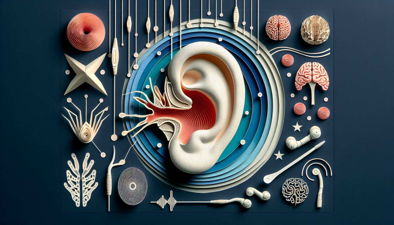
  1. Outer Ear Anatomy - The pinna and external auditory canal are like nature's megaphone, capturing sound waves and funneling them straight toward your eardrum. This design amplifies distant noises and primes them for the next adventure inside your ear. Kenhub: Auditory Pathway
  2. Middle Ear Ossicles - The malleus, incus, and stapes team up to boost tiny sound vibrations, turning whispers into signals robust enough to journey to the inner ear. It's like having a trio of mini rockstars amplifying every beat before passing the mic through the oval window. Kenhub: Auditory Pathway
  3. Cochlear Structure - The snail-shaped cochlea houses the organ of Corti, where delicate hair cells groove to the rhythm of sound waves and convert them into electrical signals. This conversion is the VIP backstage pass to your brain's auditory cortex! Wikipedia: Organ of Corti
  4. Auditory Pathway Components - Sound signals travel from the cochlear nerve through stations like the cochlear nuclei, superior olivary complex, lateral lemniscus, and inferior colliculus before reaching the medial geniculate body. Finally, the auditory cortex gets the memo and turns these electrical pulses into the melodies you perceive. TeachMeAnatomy: Auditory Pathway Guide
  5. Tonotopic Organization - The cochlea is a frequency detective, with high-pitched sounds peaking at one end of the basilar membrane and low-pitched sounds at the other. This spatial mapping makes sure your brain knows exactly which frequencies hit your eardrum. Wikipedia: Organ of Corti
  6. Sound Localization - By comparing tiny time and loudness differences of sounds arriving at each ear, the superior olivary complex acts like a sound GPS to pinpoint direction. It's how you can tell if that snack-wrapper crinkle came from the left or right! Kenhub: Auditory Pathway
  7. Inferior Colliculus Functions - This midbrain hub integrates all incoming auditory data and can trigger reflexes like turning your head toward a sudden noise. Think of it as your built-in alarm system, always on alert! Kenhub: Auditory Pathway
  8. Medial Geniculate Body Relay - Acting as the audio relay station in your thalamus, the medial geniculate body forwards processed sound signals to the auditory cortex for higher-level interpretation. Without this hand-off, you'd be stuck with raw vibrations and no symphony! Kenhub: Auditory Pathway
  9. Primary Auditory Cortex - Located on the superior temporal gyrus, this brain region decodes pitch, loudness, and location to create the sounds you recognize as speech, music, or environmental noise. It's the concert hall where your brain finally enjoys the show! Kenhub: Auditory Pathway
  10. Auditory Pathway Mnemonic (E.C.O.L.I.M.A) - Remember the sequence: Ear receptors, Cochlear nucleus, Superior Olivary nucleus, Lateral lemniscus, Inferior colliculus, Medial geniculate body, Auditory cortex. This catchy mnemonic keeps the pathway stages in perfect order so you can ace your exams! EpoMedicine: Auditory Pathway Mnemonic
Powered by: Quiz Maker