Unlock hundreds more features
Save your Quiz to the Dashboard
View and Export Results
Use AI to Create Quizzes and Analyse Results

Sign inSign in with Facebook
Sign inSign in with Google

Practice Histology Quiz: Test Tissue Knowledge

Master tissue identification, bone structure, and histology

Difficulty: Moderate
Grade: Grade 10
Study OutcomesCheat Sheet
Paper art promoting a Bone  Tissue Basics trivia quiz for high school biology students.

Which mineral is primarily responsible for the hardness of bone?
Sodium chloride
Calcium carbonate
Potassium phosphate
Hydroxyapatite
Hydroxyapatite forms the primary mineral component of bone, providing its hardness and strength. Other minerals do not contribute similarly to bone structure.
Bone is classified as which type of tissue?
Muscle tissue
Connective tissue
Nervous tissue
Epithelial tissue
Bone is a specialized connective tissue that provides structure and support to the body. It is distinct from epithelial, muscle, and nervous tissues.
Which cell is primarily responsible for the formation of new bone?
Chondrocyte
Osteocyte
Osteoblast
Osteoclast
Osteoblasts synthesize bone matrix and are essential for bone formation. The other cells serve different functions within bone or cartilage.
Which cell type is responsible for bone resorption?
Osteoblast
Osteoclast
Chondrocyte
Osteocyte
Osteoclasts are specialized cells that break down bone tissue during remodeling and repair. Other cells have roles in bone formation or maintenance.
Which type of bone tissue is denser and forms the outer layer of bones?
Spongy bone
Compact bone
Cartilaginous tissue
Periosteum
Compact bone is dense and forms the external layer of bones, providing strength and protection. Spongy bone, cartilage, and periosteum serve different roles.
What is the primary role of osteocytes in bone tissue?
Maintenance and communication within the bone matrix
Producing new bone matrix
Stimulating bone resorption
Initiating bone fractures
Osteocytes maintain bone tissue by regulating mineral content and facilitating nutrient exchange. They are not directly involved in stimulating resorption or initiating fractures.
Which process is essential for repairing a fractured bone?
Calcification
Ossification
Callus formation
Chondrification
Callus formation stabilizes a fracture by forming a temporary bone structure during healing. While ossification is part of bone development, callus formation is specifically critical for fracture repair.
What type of cells found in the periosteum are crucial for bone growth and repair?
Chondrocytes
Erythrocytes
Osteoprogenitor cells
Lymphocytes
Osteoprogenitor cells in the periosteum differentiate into osteoblasts, supporting bone growth and repair. The other cells are not involved in forming new bone tissue.
What is the primary function of the Haversian system in compact bone?
Storage of minerals
Facilitation of nerve signals
Transport of nutrients and waste
Production of bone cells
The Haversian system, or osteon, houses blood vessels that supply nutrients and remove waste from bone. It is not primarily responsible for mineral storage or nerve signaling.
Which type of cartilage is most prevalent in the embryonic skeleton and serves as a template for bone development?
Fibrocartilage
Elastic cartilage
Septal cartilage
Hyaline cartilage
Hyaline cartilage is abundant in the developing skeleton and is replaced by bone during endochondral ossification. Other types of cartilage are less common in this context.
Which ossification process involves the replacement of a cartilage template with bone?
Endochondral ossification
Intramembranous ossification
Perichondral ossification
Secondary ossification
Endochondral ossification describes the process where cartilage is gradually replaced by bone, particularly in long bones. In contrast, intramembranous ossification involves direct bone formation without a cartilage template.
How does vitamin D contribute to bone health?
Directly forms the organic bone matrix
Increases bone resorption by osteoclasts
Stimulates cartilage production
Enhances calcium absorption in the intestines
Vitamin D plays a critical role in bone health by promoting calcium absorption, which is essential for bone mineralization. It does not directly form bone matrix or increase resorption.
What is the main organic component of the bone matrix?
Elastin
Type I collagen
Fibrin
Keratin
Type I collagen is the predominant protein in the bone matrix, providing a framework for mineral deposition. The other proteins are not significant components of bone tissue.
What effect does parathyroid hormone (PTH) have on bone tissue?
Inhibits bone resorption
Promotes osteoblast differentiation exclusively
Neutralizes vitamin D
Stimulates osteoclast activity to increase blood calcium levels
Parathyroid hormone increases blood calcium levels by stimulating osteoclast-mediated bone resorption. It does not inhibit resorption or solely promote osteoblast differentiation in this process.
How does the porous structure of spongy bone benefit the skeletal system?
It enhances fat storage
It strengthens muscular attachments
It reduces bone weight while maintaining structural strength
It increases the rate of bone growth
The porous architecture of spongy bone reduces overall bone weight and provides space for bone marrow, while still contributing to structural support. It is not directly responsible for increasing growth rate or storing fat.
Which principle explains how bone structure adapts to mechanical stress, leading to changes in bone mass and architecture?
Wolff's Law
Christensen's Rule
Hardy's Principle
Mendelian Inheritance
Wolff's Law states that bone adapts to the mechanical loads placed upon it, remodeling its structure accordingly. The other options are either unrelated or not recognized in bone biology.
What sequence of events characterizes the bone healing process following a fracture?
Soft callus formation, inflammation, ossification, and complete healing
Hematoma formation, direct ossification, and immediate remodeling
Inflammation, soft callus formation, hard callus formation, and remodeling
Calcification, ossification, chondrification, and remodeling
Bone healing involves an initial inflammatory phase with hematoma formation, followed by soft callus development, hard callus formation, and finally remodeling. This sequential process is vital for effective fracture repair.
What is the significance of the lacuna-canalicular system in bone tissue?
It serves as a storage area for calcium ions
It facilitates nutrient exchange and cell communication among osteocytes
It houses blood vessels for structural support
It protects the bone from external trauma
The lacuna-canalicular system is essential for the exchange of nutrients and signaling among osteocytes. Its primary role is not to store calcium or provide direct structural support.
How do osteoblasts and osteoclasts work together during the bone remodeling process?
They coordinate in a balanced cycle, ensuring bone resorption and formation occur simultaneously
Osteoclasts transform into osteoblasts after resorption is complete
They operate independently, with osteoblasts only active during growth and osteoclasts during repair
Osteoblasts inhibit osteoclast activity to favor bone formation
Bone remodeling involves a balance between osteoclast-mediated resorption and osteoblast-mediated formation, ensuring structural integrity is maintained. This coordinated cycle is critical for overall bone health.
How does the hierarchical structure of bone contribute to its overall function in support and protection of the body?
The layered structure serves only an aesthetic purpose
The uniform density of bone prevents any uneven force distribution
The complex architecture, from microscopic collagen fibers to macroscopic bone shape, provides both strength and flexibility
The structure minimizes cellular activity, conserving energy
The hierarchical design of bone, from nanoscale collagen fibers to the overall bone shape, offers a balance of strength and flexibility that supports and protects the body. The other options do not accurately reflect the functional benefits of bone structure.
0
{"name":"Which mineral is primarily responsible for the hardness of bone?", "url":"https://www.quiz-maker.com/QPREVIEW","txt":"Which mineral is primarily responsible for the hardness of bone?, Bone is classified as which type of tissue?, Which cell is primarily responsible for the formation of new bone?","img":"https://www.quiz-maker.com/3012/images/ogquiz.png"}

Study Outcomes

  1. Understand the different types of bone tissue and their specific functions.
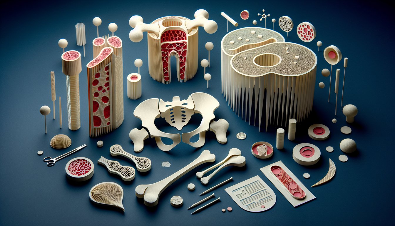
  2. Analyze the microscopic structure of bone sections to identify key features.
  3. Evaluate the relationship between tissue structure and its role in maintaining bone integrity.
  4. Apply histological concepts to interpret diagrams and models of bone and tissue.
  5. Compare and contrast various cellular structures within bone tissues.

Histology & Tissue Identification Cheat Sheet

  1. Primary tissue types - Dive into the dynamic quartet of epithelial, connective, muscle, and nervous tissues, each playing a starring role in the body's performance. Epithelial lines surfaces, connective builds scaffolds, muscle powers movement, and nervous sends the electric signals that keep you on your toes. Cement these basics with some interactive fun. Kenhub tissue quizzes
  2. Bone tissue roles - Bones are living rock stars, shaped by osteoblasts that build, osteocytes that chill in their own rooms, and osteoclasts that remodel the stage. Understanding how these cells coordinate the growth and maintenance of your skeleton is a game-changer for grasping bone health. Rock on your study with a deep-dive guide. CliffsNotes bone growth guide
  3. Histological staining - The H&E stain is like the ultimate color code for cells: hematoxylin turns nuclei blue and eosin paints cytoplasm pink, making every slide a neon map of tissue architecture. Mastering this palette helps you spot cell types and tissue patterns in milliseconds. Sharpen your skills with a comprehensive review. VAIA histology review
  4. Epithelial types - Get to know the all-star lineup: simple squamous, cuboidal, columnar, pseudostratified, stratified squamous, stratified cuboidal, and the retractable transitional epithelium. Each subtype has its favorite hangout spot, from lung air sacs to bladder walls, and unique shapes that match their jobs. Memorize their look and location to earn top marks. Quizlet histology flashcards
  5. Connective tissue composition - Think of connective tissue as the body's scaffolding, woven with collagen for tensile strength and elastin for springy resilience. Its extracellular matrix is a bustling biochemical playground that also houses cells and fibers in perfect harmony. Flip through an explainer to see the backstage mechanics. CliffsNotes connective tissue explainer
  6. Muscle tissue types - Whether you're flexing skeletal muscle, pumping blood with cardiac muscle, or digesting dinner via smooth muscle, each type has its own striation style and control system. Spotting these patterns under the microscope is like deciphering muscle Morse code. Get the scoop on each type. CliffsNotes muscle tissue breakdown
  7. Nervous tissue structure - Neurons shoot electrical messages across axons like lightning bolts, while glial cells play the VIP support crew, providing nutrients and protection. Together they form the communication superhighway that powers thoughts, movements, and reflexes. Navigate this neural network with a handy guide. CliffsNotes nervous tissue summary
  8. Microscopic practice - Hands-on experience makes perfect, so jump into histology slide quizzes to train your eye in real tissue selfies. The more slides you explore, the faster you'll recognize cellular landmarks and textural cues. Treat each quiz like a mini adventure under the lens. Kenhub slide quizzes
  9. Ossification pathways - Bones build themselves via two epic quests: intramembranous ossification straight from connective tissue, and endochondral ossification that first sculpts cartilage before hardening. Mastering these two routes reveals how your skeleton grows from embryo to adult. Plot these steps to crush your exam. CliffsNotes ossification overview
  10. Extracellular matrix magic - The ECM in connective tissues isn't just filler; it's a dynamic arena of proteins and polysaccharides that dictate tissue strength, flexibility, and cell behavior. Understanding its components and interactions unlocks insights into wound repair and disease processes. Dive into the matrix for a deeper perspective. VAIA ECM deep dive
Powered by: Quiz Maker